Imaging Modalities of the EVIS X1™ Endoscopy System: What’s Everyone Seeing?
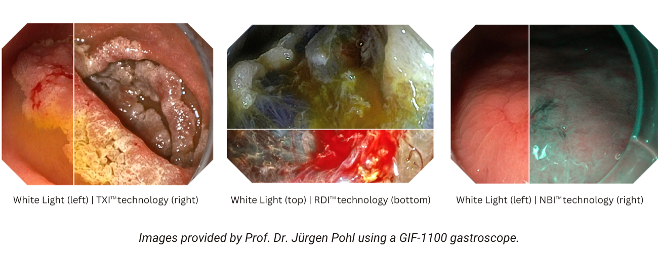
Olympus America announced the release of the EVIS X1TM endoscopy system that was unveiled in October 2023 at the American College of Gastroenterology annual meeting in Vancouver. Since then, interest has piqued in clinical data and user experience related to imaging modalities such as Red Dichromatic Imaging (RDITM) technology used to help gastroenterologists detect bleeds,1 and Narrow Band ImagingTM (NBITM) technology, used to enhance the visibility of vessels and other structures on or near the mucosal surface.1

Technological, clinical aspects of RDI technology
The best way to understand RDI imaging is to compare it to white light endoscopy (WLE) imaging. Authors of a 2022 paper published in Therapeutic Advances in Gastroenterology do just that, explaining the longer wavelength characteristics of RDI technology compared to other imaging modalities and citing use cases with potential clinical impact in the following scenarios:2
- Improving the visibility of deep blood vessels for more accurate evaluation of esophageal varices
- Detecting intraoperative bleeding during endoscopic submucosal dissection
- Evaluating disease activity in patients with ulcerative colitis
- Viewing enhanced bleeding points with the “see-through” effect, as RDI technology increases the transparency of bile and blood
- Identifying bleeding in patients with peptic ulcers and colonic diverticula
The paper also discusses the three observational modes of RDI technology available in the EVIS X1 endoscopy system, and the utility of each mode. The authors anticipate further studies to better establish the usefulness of this modality.2
RDI technology and acute GI bleeding
In a 2022 study published in Gastrointestinal Endoscopy, physicians scored visibility of the bleeding point from video footage, demonstrating the mean visibility score for all endoscopists was significantly higher in RDI (3.12 ± .51) compared with WLI (2.72 ± .50, P < 0.001). As this is a small single site retrospective study, further studies are required to validate these results.3 In an accompanying editorial, authors discussed these findings, and also noted the use of the three observational modes when using RDI technology: Mode 1 is designed for identifying bleeds, and modes 2 and 3 are designed to improve the view of deep and superficial blood vessels. The editorial posed the following questions:4
- Does better identification of bleeding points lead to shorter procedure times and improved treatment results and rebleeding rates?
- Will the use of RDI technology reduce the risk of adverse events resulting from the repeated application of cautery, especially in the duodenum or the colon?
The authors call for more research to answer these questions.
NBITM technology: Established modality, more evidence
NBI technology has had a longer run in the market, thus the evidence of its utility is better established for its prediction of several types of pathology when compared to WLE, noted David M. Poppers, MD, PhD, FACP, FASGE, AGAF, professor of medicine, division of gastroenterology at NYU Grossman School of Medicine in New York. In a May 2024 paper published in Gastroenterology & Endoscopy News, Dr. Poppers discussed the incremental advancements of NBI™ technology, and its established utility as evidenced by recommendation of its use by the American Society for Gastrointestinal Endoscopy, especially for real-time surveillance of colorectal polyps or Barrett’s esophagus.5,6 NBI technology has been shown across multiple studies to reduce procedure time by reducing the number of biopsies taken compared to the Seattle protocol for patients with Barrett’s esophagus.7 It has also been shown to increase adenoma detection rate and serrated adenoma detection rate compared to WLE.8,9
The inclusion of NBI technology in the EVIS X1™ endoscopy system marks this imaging modality’s latest iterative improvement. NBI technology on the EVIS X1 endoscopy system can be used in combination with BAI-MACTM technology, a post-processing application to increase the brightness and viewable distance of the endoscopic image. In addition to BAI-MAC technology, the EVIS X1 endoscopy system uses RDI™ technology (as discussed above) and Texture and Color Enhancement Imaging (TXITM) technology to support endoscopists to diagnose and treat GI diseases.10
To read these publications and other papers on the imaging modalities available on the EVIS X1 endoscopy system, visit our Clinical Studies page!
References
1. Data on file with Olympus (DC00489968).
2. Uraoka T, Igarashi M. Development and clinical usefulness of a unique red dichromatic imaging technology in gastrointestinal endoscopy: A narrative review. Therap Adv Gastroenterol. 2022 Sep 2;15:17562848221118302.
3. Hirai Y, Fujimoto A, Matsutani N, et al. Evaluation of the visibility of bleeding points using red dichromatic imaging in endoscopic hemostasis for acute GI bleeding (with video). Gastrointest Endosc. 2022 Apr;95(4):692-700.e3. doi: 10.1016/j.gie.2021.10.031. Epub 2021 Nov 9. PMID: 34762920.
4. Al-Sabban AHM, Al-Kawas FH. Red dichromatic imaging in acute GI bleeding: Does it make a difference? Gastrointest Endosc. 2022 Apr;95(4):701-702. doi: 10.1016/j.gie.2021.11.036. Epub 2022 Feb 2.
5. Poppers D. Advancements in GI Evaluation: Narrow Band ImagingTM Technology and the EVIS X1TM Endoscopy System. Gastroenterology & Endoscopy News. 2024 May 13(1-4).
6. ASGE Technology Committee, Abu Dayyeh BK, Thosani N, et al. ASGE Technology Committee systematic review and meta-analysis assessing the ASGE PIVI thresholds for adopting real-time endoscopic assessment of the histology of diminutive colorectal polyps. Gastrointest Endosc. 2015;81(3):502.e1-502.e16.
7. Data on file with Olympus 7/2010.
8. Atkinson NSS, Ket S, Bassett P, et al. Narrow-Band Imaging for Detection of Neoplasia at Colonoscopy: A Meta-analysis of Data From Individual Patients in Randomized Controlled Trials. Gastroenterology. 2019;157(2):462-471.
9. Aziz M, Fatima R, Lee-Smith W, et al. Comparing endoscopic interventions to improve serrated adenoma detection rates during colonoscopy: a systematic review and network meta-analysis of randomized controlled trials. Eur J Gastroenterol Hepatol. 2020;32(10):1284-1292.
10. Data on file with Olympus 6/15/2023.
The EVIS X1™ endoscopy system is not designed for cardiac applications. Other combinations of equipment may cause ventricular fibrillation or seriously affect the cardiac function of the patient. Improper use of endoscopes may result in patient injury, infection, bleeding, and/or perforation. Complete indications, contraindications, warnings, and cautions are available in the Instructions for Use (IFU).
TXI, RDI, NBI, and BAI-MAC technologies are not intended to replace histopathological sampling as a means of diagnosis. TXI, NBI, RDI, and BAI-MAC technologies are trademarks of Olympus Corporation, Olympus America, Inc., and/or their affiliates.


